Accuracy + Precision = CONFIDENCE
There is only ONE.
Find Out What's NEW!
See what leading cardiologists
are saying about HeartSee!
See the potential
Accurate detection of coronary artery disease is just the beginning. Myocardial blood flow data, when combined with perfusion imaging, can help you make important treatment decisions.1-3
In a recent prospective study, in quadrants with a significant perfusion abnormality and severely reduced coronary flow capacity (CFC), resting myocardial blood flow (MBF) increased by 11%, stress MBF increased by 59%, and the percentage of myocardium with severely reduced CFC decreased by 12% following revascularization.4
HeartSee is the innovative diagnostic tool that helps you get more from your cardiac PET imaging
- Unique coronary flow capacity (CFC) map shows regional and global coronary defects and helps categorize severity of disease.
- HeartSee delivers the CFC map in easy to interpret reports along with traditional diagnostic data.
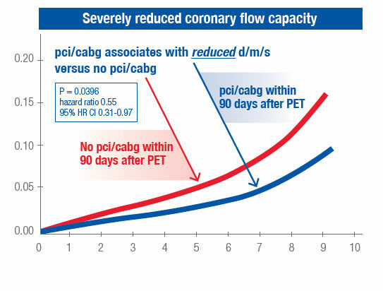
- Invasive coronary intervention within 90 days after the PET scan is associated with a 54% reduced risk of death, myocardial infarction or stroke over five years for patients who show severely reduced blood flow capacity on HeartSee’s CFC maps.3
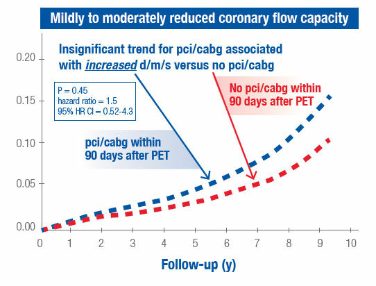
Cumulative Hazard of Death, MI and Stroke (d/m/s) Associated with Revascularization by PCI or CABG3
INDICATIONS FOR USE
HeartSee™ Software for cardiac positron emission tomography (PET) is indicated for determining regional and global absolute rest and stress myocardial perfusion in cc/min/g, Coronary Flow Reserve and their combination into the Coronary Flow Capacity (CFC) Map in patients with suspected or known coronary artery disease (CAD) in order to assist clinical interpretation of PET perfusion images by quantification of their severity.
HeartSee™ is intended for use by trained professionals such as nuclear technicians, nuclear medicine or nuclear cardiology physicians, or cardiologists with appropriate training and certification. The clinician remains ultimately responsible for the final assessment and diagnosis based on standard practices, clinical
judgment and interpretation of PET images or quantitative data.
REFERENCES
1. Gould KL, Johnson NP, MD, Bateman TM, Beanlands RS, Bengel FM, Bober R, Camici PG, Cerqueira MD, Chow BJW, Di Carli MF, Dorbala S, Gewirtz H, Gropler RJ, Kaufmann PA, Knaapen P, Juhani Knuuti J, Merhige ME, Rentrop KP, Ruddy TD, Heinrich R. Schelbert HR, Schindler TH, Schwaiger M, Sdringola S, Vitarello J, Kim Kim A. Williams Sr. KA, Gordon, D, Dilsizian V, Jagat Narula J. Anatomic Versus Physiologic Assessment of Coronary ArteryDisease, Role of Coronary Flow Reserve, Fractional Flow Reserve, and Positron Emission Tomography Imaging in Revascularization Decision-Making. JACC, Vol. 62, No. 18, 2013.
2. Bateman TM, Dilsizian V, Beanlands RS, et al. American Society of Nuclear Cardiology and Society of Nuclear Medicine and Molecular Imaging Joint Position Statement on the Clinical Indications for Myocardial Perfusion PET. J Nucl Med. 2016; 57(10):1654-1656.
3. Gould KL, Johnson NP, Roby A, Nguyen TT, Kirkeeide RL, Haynie M, Lai D, Zhu H, Patel MB, Smalling RW, Arain S, Prakash Balan P, Nguyen N, Estrera A, Sdringola S, Madjid M, Nascimbene A, Loyalka P, Kar B, Gregoric I, Safi H, McPherson D. Regional Artery Specific Thresholds of Quantitative Myocardial Perfusion by PET Associated With Reduced MI and Death After Revascularization in Stable CAD. J Nucl Med, 2018 Aug 16.
4. Bober RM, Milani RV, Ahmet A, Oktay AA, Javed F, Polin NM, Morin DP. The impact of revascularization on myocardial blood flow as assessed by positron emission tomography. European Journal of Nuclear Medicine and Molecular Imaging. Received: 18 September 2018 /Accepted: 23 January 2019.
HEARTSEE is a trademark of The University of Texas Health Science Center at Houston on behalf of the Board of Regents of The University of Texas System, and used under a license by Bracco Diagnostics Inc.


